What Are Fallen Arches
Overview

The loss of the arch of the foot (also known in some cases as the ?instep?) is called a flatfoot. People may have a very low arch or absolutely no arch whatsoever. Whilst most people with flat feet have been that way since a young age, in some people the arch height reduces over time. This can be due to systemic health concerns, degeneration of muscles and joints, hormonal changes or specific injury.
Causes
Abnormal development of the foot, producing Pes Planus, may be due to neurological problems, eg cerebral palsy, polio. Bony or ligamentous abnormalities, eg tarsal coalition (fusion of tarsal bones), accessory navicular bone. A small proportion of flexible flat feet do not correct with growth. Some of these may become rigid if the Pes Planus leads to bony changes.
Symptoms
Having flat feet can be painless and is actually normal in some people. But others with flat feet experience pain in the heel or arch area, difficulty standing on tiptoe, or have swelling along the inside of the ankle. They may also experience pain after standing for long periods of time or playing sports. Some back problems can also be attributed to flat feet.
Diagnosis
Diagnosis of flat feet or fallen arches can be made by your health practitioner and is based on the following. Clinical assessment involving visual gait assessment, as well as biomechanical assessment. A detailed family and medical history. A pain history assessment determining the location of painful symptoms. Physical palpation of the feet and painful areas. Imaging such as MRI or x-ray can be used by your practitioner to assist in the diagnosis.
arch support plantar fasciitis
Non Surgical Treatment
Treatment often consists of giving the affected foot support from underneath so that the strain is taken off it (by the use of insoles or support pads) and so symptoms are relieved. A specialist in this problem is known as a podiatrist and these do work in the NHS - ask you father's doctor whether his condition is bad enough to warrant a referral to such a specialist.
Surgical Treatment

This is rare and usually only offered if patients have significant abnormalities in their bones or muscles. Treatments include joint fusion, reshaping the bones in the foot, and occasionally moving around tendons in the foot to help balance out the stresses (called tendon transfer).
Prevention
Flat feet or Fallen Arches cannot be prevented due to congenital of nature or from underlying disease process; however, painful symptoms and future pathology from Flat Feet or Fallen Arches may be prevented by the following. Continue to wear your orthotics for work and exercise to provide stability and maintain function of your feet. Footwear. Continue to wear supportive shoes to maximise the function of your orthotic and prevent excessive movement of the joints in your feet.
After Care
Time off work depends on the type of work as well as the surgical procedures performed. . A patient will be required to be non-weight bearing in a cast or splint and use crutches for four to twelve weeks. Usually a patient can return to work in one to two weeks if they are able to work while seated. If a person's job requires standing and walking, return to work may take several weeks. Complete recovery may take six months to a full year. Complications can occur as with all surgeries, but are minimized by strictly following your surgeon's post-operative instructions. The main complications include infection, bone that is slow to heal or does not heal, progression or reoccurrence of deformity, a stiff foot, and the need for further surgery. Many of the above complications can be avoided by only putting weight on the operative foot when allowed by your surgeon.
The loss of the arch of the foot (also known in some cases as the ?instep?) is called a flatfoot. People may have a very low arch or absolutely no arch whatsoever. Whilst most people with flat feet have been that way since a young age, in some people the arch height reduces over time. This can be due to systemic health concerns, degeneration of muscles and joints, hormonal changes or specific injury.
Causes
Abnormal development of the foot, producing Pes Planus, may be due to neurological problems, eg cerebral palsy, polio. Bony or ligamentous abnormalities, eg tarsal coalition (fusion of tarsal bones), accessory navicular bone. A small proportion of flexible flat feet do not correct with growth. Some of these may become rigid if the Pes Planus leads to bony changes.
Symptoms
Having flat feet can be painless and is actually normal in some people. But others with flat feet experience pain in the heel or arch area, difficulty standing on tiptoe, or have swelling along the inside of the ankle. They may also experience pain after standing for long periods of time or playing sports. Some back problems can also be attributed to flat feet.
Diagnosis
Diagnosis of flat feet or fallen arches can be made by your health practitioner and is based on the following. Clinical assessment involving visual gait assessment, as well as biomechanical assessment. A detailed family and medical history. A pain history assessment determining the location of painful symptoms. Physical palpation of the feet and painful areas. Imaging such as MRI or x-ray can be used by your practitioner to assist in the diagnosis.
arch support plantar fasciitis
Non Surgical Treatment
Treatment often consists of giving the affected foot support from underneath so that the strain is taken off it (by the use of insoles or support pads) and so symptoms are relieved. A specialist in this problem is known as a podiatrist and these do work in the NHS - ask you father's doctor whether his condition is bad enough to warrant a referral to such a specialist.
Surgical Treatment
This is rare and usually only offered if patients have significant abnormalities in their bones or muscles. Treatments include joint fusion, reshaping the bones in the foot, and occasionally moving around tendons in the foot to help balance out the stresses (called tendon transfer).
Prevention
Flat feet or Fallen Arches cannot be prevented due to congenital of nature or from underlying disease process; however, painful symptoms and future pathology from Flat Feet or Fallen Arches may be prevented by the following. Continue to wear your orthotics for work and exercise to provide stability and maintain function of your feet. Footwear. Continue to wear supportive shoes to maximise the function of your orthotic and prevent excessive movement of the joints in your feet.
After Care
Time off work depends on the type of work as well as the surgical procedures performed. . A patient will be required to be non-weight bearing in a cast or splint and use crutches for four to twelve weeks. Usually a patient can return to work in one to two weeks if they are able to work while seated. If a person's job requires standing and walking, return to work may take several weeks. Complete recovery may take six months to a full year. Complications can occur as with all surgeries, but are minimized by strictly following your surgeon's post-operative instructions. The main complications include infection, bone that is slow to heal or does not heal, progression or reoccurrence of deformity, a stiff foot, and the need for further surgery. Many of the above complications can be avoided by only putting weight on the operative foot when allowed by your surgeon.
Measuring Apparent Leg Length Discrepancy
Overview
leg length discrepancy can be present from birth (congenital) or acquired (a result of an injury, infection or tumor). Some of the conditions that can cause limb problems in a child or young adult include congenital conditions present from birth. Osteogenesis imperfecta,. Bow legs. Knock knees. Neurofibromatosis. Arthritis. Infections of the bones and tumors. Injuries involving the growth center of the bone. There may also be deformities that are a result of the soft tissues and not the bones, such as with arthrogryposis and burns.
Causes
Leg length discrepancies can be caused by: hip and knee replacements, lower limb injuries, bone diseases, neuromuscular issues and congenital problems. Although discrepancies of 2 cm or less are most common, discrepancies can be greater than 6 cm. People who have LLD tend to make up for the difference by over bending their longer leg or standing on the toes of their shorter leg. This compensation leads to an inefficient, up and down gait, which is quite tiring and over time can result in posture problems as well as pain in the back, hips, knees and ankles.
Symptoms
As patients develop LLD, they will naturally and even unknowingly attempt to compensate for the difference between their two legs by either bending the longer leg excessively or standing on the toes of the short leg. When walking, they are forced to step down on one side and thrust upwards on the other side, which leads to a gait pattern with an abnormal up and down motion. For many patients, especially adolescents, the appearance of their gait may be more personally troublesome than any symptoms that arise or any true functional deficiency. Over time, standing on one's toes can create a contracture at the ankle, in which the calf muscle becomes abnormally contracted, a condition that can help an LLD patient with walking, but may later require surgical repair. If substantial enough, LLD left untreated can contribute to other serious orthopaedic problems, such as degenerative arthritis, scoliosis, or lower back pain. However, with proper treatment, children with leg length discrepancy generally do quite well, without lingering functional or cosmetic deficiencies.
Diagnosis
The evaluation of leg length discrepancy typically involves sequential x-rays to measure the exact discrepancy, while following its progression. In addition, an x-ray of the wrist allows us to more carefully age your child. Skeletal age and chronological age do not necessarily equal each other and frequently a child's bone age will be significantly different than his or her stated age. Your child's physician can establish a treatment plan once all the facts are known: the bone age, the exact amount of discrepancy, and the cause, if it can be identified.
Non Surgical Treatment
Treatment for an LLD depends on the amount of difference and the cause, if known. The doctor will discuss treatment options carefully with you and your child before any decisions are made. It is important to note that treatment is planned with the child?s final height and leg lengths in mind, not the current leg lengths. Treatment is generally not needed if the child?s final LLD is predicted to be 2 centimeters or less at full height. However, the child should return to an orthopaedic doctor by age 10 for re-evaluation. Treatment is often recommended for LLDs predicted to be more than 2 centimeters at full height. If treatment is done, it usually doesn?t begin until the child starts walking. Possible treatment options include, A ?lift? in one shoe to level the child?s hips. This is often the only treatment needed for small discrepancies.

what are shoe lifts?
Surgical Treatment
The bone is lengthened by surgically applying an external fixation device to the leg. The external fixator, a scaffold-like frame, is connected to the bone with wires, pins, or both. A small crack is made in the bone and the frame creates tension when the patient or family member turns its dial. This is done several times each day. The lengthening process begins approximately five to 10 days after surgery. The bone may lengthen 1 millimeter per day, or approximately 1 inch per month. Lengthening may be slower in a bone that was previously injured. It may also be slower if the leg was operated on before. Bones in patients with potential blood vessel abnormalities, such as cigarette smokers, may also need to be lengthened more slowly. The external fixator is worn until the bone is strong enough to support the patient safely. This usually takes about three months for each inch. Factors such as age, health, smoking and participation in rehabilitation can affect the amount of time needed.
leg length discrepancy can be present from birth (congenital) or acquired (a result of an injury, infection or tumor). Some of the conditions that can cause limb problems in a child or young adult include congenital conditions present from birth. Osteogenesis imperfecta,. Bow legs. Knock knees. Neurofibromatosis. Arthritis. Infections of the bones and tumors. Injuries involving the growth center of the bone. There may also be deformities that are a result of the soft tissues and not the bones, such as with arthrogryposis and burns.
Causes
Leg length discrepancies can be caused by: hip and knee replacements, lower limb injuries, bone diseases, neuromuscular issues and congenital problems. Although discrepancies of 2 cm or less are most common, discrepancies can be greater than 6 cm. People who have LLD tend to make up for the difference by over bending their longer leg or standing on the toes of their shorter leg. This compensation leads to an inefficient, up and down gait, which is quite tiring and over time can result in posture problems as well as pain in the back, hips, knees and ankles.
Symptoms
As patients develop LLD, they will naturally and even unknowingly attempt to compensate for the difference between their two legs by either bending the longer leg excessively or standing on the toes of the short leg. When walking, they are forced to step down on one side and thrust upwards on the other side, which leads to a gait pattern with an abnormal up and down motion. For many patients, especially adolescents, the appearance of their gait may be more personally troublesome than any symptoms that arise or any true functional deficiency. Over time, standing on one's toes can create a contracture at the ankle, in which the calf muscle becomes abnormally contracted, a condition that can help an LLD patient with walking, but may later require surgical repair. If substantial enough, LLD left untreated can contribute to other serious orthopaedic problems, such as degenerative arthritis, scoliosis, or lower back pain. However, with proper treatment, children with leg length discrepancy generally do quite well, without lingering functional or cosmetic deficiencies.
Diagnosis
The evaluation of leg length discrepancy typically involves sequential x-rays to measure the exact discrepancy, while following its progression. In addition, an x-ray of the wrist allows us to more carefully age your child. Skeletal age and chronological age do not necessarily equal each other and frequently a child's bone age will be significantly different than his or her stated age. Your child's physician can establish a treatment plan once all the facts are known: the bone age, the exact amount of discrepancy, and the cause, if it can be identified.
Non Surgical Treatment
Treatment for an LLD depends on the amount of difference and the cause, if known. The doctor will discuss treatment options carefully with you and your child before any decisions are made. It is important to note that treatment is planned with the child?s final height and leg lengths in mind, not the current leg lengths. Treatment is generally not needed if the child?s final LLD is predicted to be 2 centimeters or less at full height. However, the child should return to an orthopaedic doctor by age 10 for re-evaluation. Treatment is often recommended for LLDs predicted to be more than 2 centimeters at full height. If treatment is done, it usually doesn?t begin until the child starts walking. Possible treatment options include, A ?lift? in one shoe to level the child?s hips. This is often the only treatment needed for small discrepancies.
what are shoe lifts?
Surgical Treatment
The bone is lengthened by surgically applying an external fixation device to the leg. The external fixator, a scaffold-like frame, is connected to the bone with wires, pins, or both. A small crack is made in the bone and the frame creates tension when the patient or family member turns its dial. This is done several times each day. The lengthening process begins approximately five to 10 days after surgery. The bone may lengthen 1 millimeter per day, or approximately 1 inch per month. Lengthening may be slower in a bone that was previously injured. It may also be slower if the leg was operated on before. Bones in patients with potential blood vessel abnormalities, such as cigarette smokers, may also need to be lengthened more slowly. The external fixator is worn until the bone is strong enough to support the patient safely. This usually takes about three months for each inch. Factors such as age, health, smoking and participation in rehabilitation can affect the amount of time needed.
Heel Pain And Discomfort
Overview

Heel pain is a very challenging problem as it can be local and/or referred. It has been more prevalent recently due to the hard grounds on which people have to run. Commonly people will present with heel pain, thrusting an x-ray at you, and being adamant that the problem is a ?heel spur?. This is defined as a small bone that grows from the heel, directing forwards towards the toes. This may be as small as 1 mm to anything as large as 8 - 10 mm. Most of the time, this is an incidental finding, as there many heels that are pain free that have heel spurs evident on x-rays. The spur is thought to be a result of traction of the plantar fascia on the heel. In some cases, the spur may contribute to the symptoms, but is not the main cause. This should be explained very carefully to the patient, as the focus on the spur may limit the recovery, as the patient may believe that the only way to eliminate the pain is to remove the spur.
Causes
While heel pain has many causes, it is usually the result of poor biomechanics (abnormalities in the way we walk). This can place too much stress on the heel bone and the soft tissues attached to it. The stress may result from injury, or a bruise incurred while walking, running or jumping on hard surfaces: wearing poorly constructed footwear or being significantly over weight. Systemic diseases such as arthritis can also contribute to heel pain.
Symptoms
Both heel pain and heel spurs are frequently associated with an inflammation of the long band of tissue that connects the heel and the ball of the foot. The inflammation of this arch area is called plantar fasciitis. The inflammation maybe aggravated by shoes that lack appropriate support and by the chronic irritation that sometimes accompanies an athletic lifestyle. Achilles Tendinopathy, Pain and inflammation of the tendon at the back of the heel that connects the calf muscle to the foot. Sever?s, Often found in children between the ages of 8 - 13 years and is an inflammation of the calcaneal epiphyseal plate (growth plate) in the back of the heel. Bursitis, An inflamed bursa is a small irritated sack of fluid at the back of the heel. Other types of heel pain include soft tissue growths, Haglunds deformity (bone enlargement at the back of the heel), bruises or stress fractures and possible nerve entrapment.
Diagnosis
After you have described your foot symptoms, your doctor will want to know more details about your pain, your medical history and lifestyle, including. Whether your pain is worse at specific times of the day or after specific activities. Any recent injury to the area. Your medical and orthopedic history, especially any history of diabetes, arthritis or injury to your foot or leg. Your age and occupation. Your recreational activities, including sports and exercise programs. The type of shoes you usually wear, how well they fit, and how frequently you buy a new pair. Your doctor will examine you, including. An evaluation of your gait. While you are barefoot, your doctor will ask you to stand still and to walk in order to evaluate how your foot moves as you walk. An examination of your feet. Your doctor may compare your feet for any differences between them. Then your doctor may examine your painful foot for signs of tenderness, swelling, discoloration, muscle weakness and decreased range of motion. A neurological examination. The nerves and muscles may be evaluated by checking strength, sensation and reflexes. In addition to examining you, your health care professional may want to examine your shoes. Signs of excessive wear in certain parts of a shoe can provide valuable clues to problems in the way you walk and poor bone alignment. Depending on the results of your physical examination, you may need foot X-rays or other diagnostic tests.
Non Surgical Treatment
Treatment of heel pain generally occurs in stages. At the earliest sign of heel pain, aggressive calf muscle stretching should be started. Additionally, taking an oral anti-inflammatory medication and over-the- counter arch supports or heel cushions may be beneficial. The next phase of treatment might consist of continued calf muscle stretching exercises, cortisone injections and orthopedic taping of the foot to support the arch. If this treatment fails, or if there is reoccurrence of the heel pain, then functional foot orthotics might be considered. A functional orthotic is a device that is prescribed and fitted by your foot doctor, which fits in normal shoes like an arch support. Unlike an arch support, however the orthotic corrects abnormal pronation of the subtalar joint. Thus orthotics address the cause of the heel pain - abnormal pronation of the foot. Pump bump, treatment is similar to the treatment of bursitis and heel spurs. In rare cases, the bony growth at the heel may need to be removed surgically. Heel bruises can be treated by applying an ice pack for the first few minutes after injury. Achilles tendonitis, this condition is treated conservatively with rest, NSAIDs and physical therapy. If a sprain, fracture or other injury has caused the trapped nerve, this underlying problem must be treated first. In rare cases, surgery may be done to release the trapped nerve.
Surgical Treatment
Surgery to correct heel pain is generally only recommended if orthotic treatment has failed. There are some exceptions to this course of treatment and it is up to you and your doctor to determine the most appropriate course of treatment. Following surgical treatment to correct heel pain the patient will generally have to continue the use of orthotics. The surgery does not correct the cause of the heel pain. The surgery will eliminate the pain but the process that caused the pain will continue without the use of orthotics. If orthotics have been prescribed prior to surgery they generally do not have to be remade.
deelsonheels
Prevention

Flexibility is key when it comes to staving off the pain associated with these heel conditions. The body is designed to work in harmony, so stretching shouldn?t be concentrated solely on the foot itself. The sympathetic tendons and muscles that move the foot should also be stretched and gently exercised to ensure the best results for your heel stretches. Take the time to stretch thighs, calves and ankles to encourage healthy blood flow and relaxed muscle tension that will keep pain to a minimum. If ice is recommended by a doctor, try freezing a half bottle of water and slowly rolling your bare foot back and forth over it for as long as is comfortable. The use of elastic or canvas straps to facilitate stretching of an extended leg can also be helpful when stretching without an assistant handy. Once cleared by a doctor, a daily regimen of over-the-counter anti-inflammatory medication like Naproxen Sodium will keep pain at bay and increase flexibility in those afflicted by heel pain. While this medication is not intended to act as a substitute for medical assessments, orthopedics or stretching, it can nonetheless be helpful in keeping discomfort muted enough to enjoy daily life. When taking any medication for your heel pain, be sure to follow directions regarding food and drink, and ask your pharmacist about possible interactions with existing medications or frequent activities.
Heel pain is a very challenging problem as it can be local and/or referred. It has been more prevalent recently due to the hard grounds on which people have to run. Commonly people will present with heel pain, thrusting an x-ray at you, and being adamant that the problem is a ?heel spur?. This is defined as a small bone that grows from the heel, directing forwards towards the toes. This may be as small as 1 mm to anything as large as 8 - 10 mm. Most of the time, this is an incidental finding, as there many heels that are pain free that have heel spurs evident on x-rays. The spur is thought to be a result of traction of the plantar fascia on the heel. In some cases, the spur may contribute to the symptoms, but is not the main cause. This should be explained very carefully to the patient, as the focus on the spur may limit the recovery, as the patient may believe that the only way to eliminate the pain is to remove the spur.
Causes
While heel pain has many causes, it is usually the result of poor biomechanics (abnormalities in the way we walk). This can place too much stress on the heel bone and the soft tissues attached to it. The stress may result from injury, or a bruise incurred while walking, running or jumping on hard surfaces: wearing poorly constructed footwear or being significantly over weight. Systemic diseases such as arthritis can also contribute to heel pain.
Symptoms
Both heel pain and heel spurs are frequently associated with an inflammation of the long band of tissue that connects the heel and the ball of the foot. The inflammation of this arch area is called plantar fasciitis. The inflammation maybe aggravated by shoes that lack appropriate support and by the chronic irritation that sometimes accompanies an athletic lifestyle. Achilles Tendinopathy, Pain and inflammation of the tendon at the back of the heel that connects the calf muscle to the foot. Sever?s, Often found in children between the ages of 8 - 13 years and is an inflammation of the calcaneal epiphyseal plate (growth plate) in the back of the heel. Bursitis, An inflamed bursa is a small irritated sack of fluid at the back of the heel. Other types of heel pain include soft tissue growths, Haglunds deformity (bone enlargement at the back of the heel), bruises or stress fractures and possible nerve entrapment.
Diagnosis
After you have described your foot symptoms, your doctor will want to know more details about your pain, your medical history and lifestyle, including. Whether your pain is worse at specific times of the day or after specific activities. Any recent injury to the area. Your medical and orthopedic history, especially any history of diabetes, arthritis or injury to your foot or leg. Your age and occupation. Your recreational activities, including sports and exercise programs. The type of shoes you usually wear, how well they fit, and how frequently you buy a new pair. Your doctor will examine you, including. An evaluation of your gait. While you are barefoot, your doctor will ask you to stand still and to walk in order to evaluate how your foot moves as you walk. An examination of your feet. Your doctor may compare your feet for any differences between them. Then your doctor may examine your painful foot for signs of tenderness, swelling, discoloration, muscle weakness and decreased range of motion. A neurological examination. The nerves and muscles may be evaluated by checking strength, sensation and reflexes. In addition to examining you, your health care professional may want to examine your shoes. Signs of excessive wear in certain parts of a shoe can provide valuable clues to problems in the way you walk and poor bone alignment. Depending on the results of your physical examination, you may need foot X-rays or other diagnostic tests.
Non Surgical Treatment
Treatment of heel pain generally occurs in stages. At the earliest sign of heel pain, aggressive calf muscle stretching should be started. Additionally, taking an oral anti-inflammatory medication and over-the- counter arch supports or heel cushions may be beneficial. The next phase of treatment might consist of continued calf muscle stretching exercises, cortisone injections and orthopedic taping of the foot to support the arch. If this treatment fails, or if there is reoccurrence of the heel pain, then functional foot orthotics might be considered. A functional orthotic is a device that is prescribed and fitted by your foot doctor, which fits in normal shoes like an arch support. Unlike an arch support, however the orthotic corrects abnormal pronation of the subtalar joint. Thus orthotics address the cause of the heel pain - abnormal pronation of the foot. Pump bump, treatment is similar to the treatment of bursitis and heel spurs. In rare cases, the bony growth at the heel may need to be removed surgically. Heel bruises can be treated by applying an ice pack for the first few minutes after injury. Achilles tendonitis, this condition is treated conservatively with rest, NSAIDs and physical therapy. If a sprain, fracture or other injury has caused the trapped nerve, this underlying problem must be treated first. In rare cases, surgery may be done to release the trapped nerve.
Surgical Treatment
Surgery to correct heel pain is generally only recommended if orthotic treatment has failed. There are some exceptions to this course of treatment and it is up to you and your doctor to determine the most appropriate course of treatment. Following surgical treatment to correct heel pain the patient will generally have to continue the use of orthotics. The surgery does not correct the cause of the heel pain. The surgery will eliminate the pain but the process that caused the pain will continue without the use of orthotics. If orthotics have been prescribed prior to surgery they generally do not have to be remade.
deelsonheels
Prevention
Flexibility is key when it comes to staving off the pain associated with these heel conditions. The body is designed to work in harmony, so stretching shouldn?t be concentrated solely on the foot itself. The sympathetic tendons and muscles that move the foot should also be stretched and gently exercised to ensure the best results for your heel stretches. Take the time to stretch thighs, calves and ankles to encourage healthy blood flow and relaxed muscle tension that will keep pain to a minimum. If ice is recommended by a doctor, try freezing a half bottle of water and slowly rolling your bare foot back and forth over it for as long as is comfortable. The use of elastic or canvas straps to facilitate stretching of an extended leg can also be helpful when stretching without an assistant handy. Once cleared by a doctor, a daily regimen of over-the-counter anti-inflammatory medication like Naproxen Sodium will keep pain at bay and increase flexibility in those afflicted by heel pain. While this medication is not intended to act as a substitute for medical assessments, orthopedics or stretching, it can nonetheless be helpful in keeping discomfort muted enough to enjoy daily life. When taking any medication for your heel pain, be sure to follow directions regarding food and drink, and ask your pharmacist about possible interactions with existing medications or frequent activities.
Managing Mortons Neuroma
Overview
 Mortons Neuroma is a common painful condition involving compression of nerves between the long bones of the forefoot just before they enter the toes. Commonly this involves the 3rd and 4th toes, however may affect the 2nd and 3rd toes. Repeated trauma or compression of these nerves causes the nerves to swell and thicken causing a Morton's neuroma to develop.
Causes
Inappropriate footwear is one of the principle causes of Morton?s neuroma. Toe spring and tapering toe boxes are the most problematic shoe design features that contribute to this health problem. Morton?s neuroma occurs when one of your nerves is stretched and pinched, which happens with great frequency in people who wear shoes incorporating these design features. A professional shoe fitting should always be sought if you are struggling with neuroma-related symptoms.
Symptoms
Symptoms include tingling in the space between the third and fourth toes, toe cramping, a sharp, shooting, or burning pain in the ball of the foot and sometimes toes, pain that increases when wearing shoes or pressing on the area, pain that gets worse over time. In rare cases, nerve pain occurs in the space between the second and third toes. This is not a common form of Morton neuroma, but treatment is similar.
Diagnosis
If you suspect Morton?s Neuroma, it is essential that you confirm your suspicions by consulting with a podiatric physician. Don?t wait for the symptoms to go away for good (even if they tend to come and go). Also, remember that many conditions have similar symptoms, and only a professional can tell the difference.
Non Surgical Treatment
Treaments may include wearing wider shoes to reduce the squeezing force on the foot. Adding a specially made padding to shoes to offload the pressure on the ball of the foot (called a metatarsal dome) Addressing the foot and lower limb biomechanics. This involves looking at foot stability and if needed prescribing an orthotic device to correct your foot position. Anesthetic & Cortisone injections. This is done when the above treatments are insufficient. The trauma is sometimes so great that conservative treatment cannot control the inflammation or cause of the pain. A series of injections are performed to control the inflammation or to temporarily settle the nerve. An ultrasound and cortisone injection can be prescribed by your podiatrist.
Surgical Treatment
Surgery is occasionally required when the conservative treatment is not able to relieve your symptoms, particularly if you have had pain for more than 6 months. 80% of patients who require surgery report good results, with 71% of people becoming pain-free.
Mortons Neuroma is a common painful condition involving compression of nerves between the long bones of the forefoot just before they enter the toes. Commonly this involves the 3rd and 4th toes, however may affect the 2nd and 3rd toes. Repeated trauma or compression of these nerves causes the nerves to swell and thicken causing a Morton's neuroma to develop.Causes
Inappropriate footwear is one of the principle causes of Morton?s neuroma. Toe spring and tapering toe boxes are the most problematic shoe design features that contribute to this health problem. Morton?s neuroma occurs when one of your nerves is stretched and pinched, which happens with great frequency in people who wear shoes incorporating these design features. A professional shoe fitting should always be sought if you are struggling with neuroma-related symptoms.
Symptoms
Symptoms include tingling in the space between the third and fourth toes, toe cramping, a sharp, shooting, or burning pain in the ball of the foot and sometimes toes, pain that increases when wearing shoes or pressing on the area, pain that gets worse over time. In rare cases, nerve pain occurs in the space between the second and third toes. This is not a common form of Morton neuroma, but treatment is similar.
Diagnosis
If you suspect Morton?s Neuroma, it is essential that you confirm your suspicions by consulting with a podiatric physician. Don?t wait for the symptoms to go away for good (even if they tend to come and go). Also, remember that many conditions have similar symptoms, and only a professional can tell the difference.
Non Surgical Treatment
Treaments may include wearing wider shoes to reduce the squeezing force on the foot. Adding a specially made padding to shoes to offload the pressure on the ball of the foot (called a metatarsal dome) Addressing the foot and lower limb biomechanics. This involves looking at foot stability and if needed prescribing an orthotic device to correct your foot position. Anesthetic & Cortisone injections. This is done when the above treatments are insufficient. The trauma is sometimes so great that conservative treatment cannot control the inflammation or cause of the pain. A series of injections are performed to control the inflammation or to temporarily settle the nerve. An ultrasound and cortisone injection can be prescribed by your podiatrist.
Surgical Treatment
Surgery is occasionally required when the conservative treatment is not able to relieve your symptoms, particularly if you have had pain for more than 6 months. 80% of patients who require surgery report good results, with 71% of people becoming pain-free.
How Shoe Lifts Remedy Leg Length Difference
There are actually not one but two different types of leg length discrepancies, congenital and acquired. Congenital indicates that you are born with it. One leg is structurally shorter in comparison to the other. Through developmental phases of aging, the brain picks up on the walking pattern and recognizes some difference. The human body typically adapts by dipping one shoulder to the "short" side. A difference of under a quarter inch is not very uncommon, doesn't need Shoe Lifts to compensate and ordinarily doesn't have a profound effect over a lifetime.

Leg length inequality goes mainly undiagnosed on a daily basis, however this condition is simply fixed, and can reduce many instances of low back pain.
Treatment for leg length inequality typically consists of Shoe Lifts. These are low-priced, often being under twenty dollars, compared to a custom orthotic of $200 plus. When the amount of leg length inequality begins to exceed half an inch, a whole sole lift is generally the better choice than a heel lift. This prevents the foot from being unnecessarily stressed in an abnormal position.
Chronic back pain is the most common ailment afflicting people today. Over 80 million men and women are affected by back pain at some stage in their life. It's a problem that costs companies vast amounts of money every year as a result of lost time and production. Innovative and superior treatment solutions are always sought after in the hope of minimizing the economical influence this condition causes.

Men and women from all corners of the world suffer the pain of foot ache due to leg length discrepancy. In these cases Shoe Lifts can be of beneficial. The lifts are capable of eliminating any pain and discomfort in the feet. Shoe Lifts are recommended by countless qualified orthopaedic physicians.
In order to support the body in a well-balanced fashion, feet have a crucial function to play. In spite of that, it is sometimes the most overlooked area in the human body. Many people have flat-feet which means there may be unequal force placed on the feet. This causes other areas of the body including knees, ankles and backs to be affected too. Shoe Lifts guarantee that the right posture and balance are restored.
Leg length inequality goes mainly undiagnosed on a daily basis, however this condition is simply fixed, and can reduce many instances of low back pain.
Treatment for leg length inequality typically consists of Shoe Lifts. These are low-priced, often being under twenty dollars, compared to a custom orthotic of $200 plus. When the amount of leg length inequality begins to exceed half an inch, a whole sole lift is generally the better choice than a heel lift. This prevents the foot from being unnecessarily stressed in an abnormal position.
Chronic back pain is the most common ailment afflicting people today. Over 80 million men and women are affected by back pain at some stage in their life. It's a problem that costs companies vast amounts of money every year as a result of lost time and production. Innovative and superior treatment solutions are always sought after in the hope of minimizing the economical influence this condition causes.
Men and women from all corners of the world suffer the pain of foot ache due to leg length discrepancy. In these cases Shoe Lifts can be of beneficial. The lifts are capable of eliminating any pain and discomfort in the feet. Shoe Lifts are recommended by countless qualified orthopaedic physicians.
In order to support the body in a well-balanced fashion, feet have a crucial function to play. In spite of that, it is sometimes the most overlooked area in the human body. Many people have flat-feet which means there may be unequal force placed on the feet. This causes other areas of the body including knees, ankles and backs to be affected too. Shoe Lifts guarantee that the right posture and balance are restored.
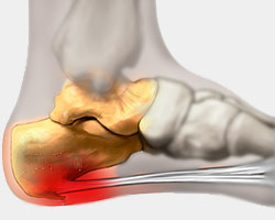
Posterior Calcaneal Spur Causes

Overview
Heel spurs are usually under the heel and are generally caused by excessive forces acting on the bone. By far the most common cause of heel spurs is abnormal biomechanics - often the same biomechanics that cause plantar fasciitis. Heel spurs are not a direct cause of heel pain. They grow in response to the forces of the soft tissue pulling on the bone. Any condition where the foot has excessive motion can produce tension within the soft tissues acting on the heel.
Causes
Heel spurs form in some patients who have plantar fasciitis (PLAN-tar fash-ee-I-tis), and tend to occur in patients who have had the problem for a prolonged period of time. While about 70 percent of patients with plantar fasciitis have a heel spur, X-rays also show about 50 percent of patients with no symptoms of plantar fasciitis also have a heel spur.

Symptoms
Bone spurs may cause sudden, severe pain when putting weight on the affected foot. Individuals may try to walk on their toes or ball of the foot to avoid painful pressure on the heel spur. This compensation during walking or running can cause additional problems in the ankle, knee, hip, or back.
Diagnosis
Your doctor will review your medical history and examine your foot. X-rays are used to identify the location and size of the heel spur.
Non Surgical Treatment
In many cases treatment is non-surgical and can relieve pain, but may take from three months to a year to fully recover. Performing stretching exercises to help relax the tissues in the heel as well as rest, icing, and over-the-counter anti-inflammatory or prescription medications can help ease symptoms. Customized orthotics or shoe inserts to position and cushion your heel can help.
Surgical Treatment
When chronic heel pain fails to respond to conservative treatment, surgical treatment may be necessary. Heel surgery can provide relief of pain and restore mobility. The type of procedure used is based on examination and usually consists of releasing the excessive tightness of the plantar fascia, called a plantar fascia release. Depending on the presence of excess bony build up, the procedure may or may not include removal of heel spurs. Similar to other surgical interventions, there are various modifications and surgical enhancements regarding surgery of the heel.
Prevention
If you have not yet developed this condition, you can take steps to protect yourself from it. Most importantly, make it a rule to wear properly fitted footwear. Avoid shoes that have become worn down in the heel, and don't choose shoes that cause you to walk in an abnormal fashion. Maintaining a healthy weight will ensure that undue pressure isn't being put on the ligaments, tendons and bones of your feet. If your job requires a great deal of time on your feet, or if you exercise regularly, be sure to balance periods of activity with periods of rest for your feet.
Controlling Posterior Calcaneal Spur
Overview
Heel spurs are a condition that usually makes its presence known first thing in the morning via heel pain. Discomfort is typically felt in the front and bottom of the heel (calcaneal). Pain can be constant for several months or intermittent for lengthy periods of time.
Causes
Though this syndrome is most common in individuals 40 years or older, it can occur at any age. The following factors increase the likelihood of heel spur development. An uneven gait which applies too much pressure to certain areas of the foot. Being overweight. Wearing worn shoes or ill-fitting footwear. Job conditions that require long periods spent standing or lifting heavy objects. The normal aging process which results in a decrease in ligament elasticity.

Symptoms
The Heel Spur itself is not thought to be painful. Patients who experience pain with Plantar Fasciitis are suffering from inflammation and irritation of the plantar fascia. This the primary cause of pain and not the Heel Spur. Heel Spurs form in some patients who have plantar fasciitis, and tend to occur in patients who have had the problem for a prolonged period of time. While about 70 % of patients with plantar fasciitis have a heel spur, X-rays also show about 50 % of patients with no symptoms of plantar fasciitis also have a heel spur.
Diagnosis
Diagnosis of a heel spur can be done with an x-ray, which will be able to reveal the bony spur. Normally, it occurs where the plantar fascia connects to the heel bone. When the plantar fascia ligament is pulled excessively it begins to pull away from the heel bone. When this excessive pulling occurs, it causes the body to respond by depositing calcium in the injured area, resulting in the formation of the bone spur. The Plantar fascia ligament is a fibrous band of connective tissue running between the heel bone and the ball of the foot. This structure maintains the arch of the foot and distributes weight along the foot as we walk. However, due to the stress that this ligament must endure, it can easily become damaged which commonly occurs along with heel spurs.
Non Surgical Treatment
If pain and other symptoms of inflammation-redness, swelling, heat-persist, you should limit normal daily activities and contact a doctor of podiatric medicine. The podiatric physician will examine the area and may perform diagnostic X-rays to rule out problems of the bone. Early treatment might involve oral or injectable anti-inflammatory medication, exercise and shoe recommendations, taping or strapping, or use of shoe inserts or orthotic devices. Taping or strapping supports the foot, placing stressed muscles and tendons in a physiologically restful state. Physical therapy may be used in conjunction with such treatments. A functional orthotic device may be prescribed for correcting biomechanical imbalance, controlling excessive pronation, and supporting of the ligaments and tendons attaching to the heel bone. It will effectively treat the majority of heel and arch pain without the need for surgery. Only a relatively few cases of heel pain require more advanced treatments or surgery. If surgery is necessary, it may involve the release of the plantar fascia, removal of a spur, removal of a bursa, or removal of a neuroma or other soft-tissue growth.
Surgical Treatment
In a small number of cases (usually less than 5 percent), patients may not experience relief after trying the recommendations listed above. It is important that conservative treatments (such as those listed above) be performed for AT LEAST a year before considering surgery. Time is important in curing the pain from heel spurs, and insufficient treatment before surgery may subject you to potential complications from the procedure. If these treatments fail, your doctor may consider an operation to loosen the plantar fascia, called a plantar fascia release.